Loculated Pleural Effusion Differential Diagnosis / Transcatheter Intrapleural Urokinase For Loculated Pleural Effusion. Pleural effusion is suspected in patients with pleuritic pain, unexplained dyspnea, or suggestive signs. Loculated right pleural effusion with foci of atelectasis and consolidative changes concerning for pneumonia. Clinical manifestations include chest pain, cough, and dyspnea. Congestive heart failure (most common) Pleural effusions can loculate as a result of adhesions.
Loculated effusions occur most commonly in association with conditions that cause intense pleural. Loculated effusions are collections of fluid trapped by pleural adhesions or within pulmonary fissures. Diffuse nodules and opacification in right lung with compressive atelectasis. 8 pleural effusion differential diagnosis. Pleural effusion is suspected in patients with pleuritic pain, unexplained dyspnea, or suggestive signs.
Tuberculous Pleural Effusion Shaw 2019 Respirology Wiley Online Library from onlinelibrary.wiley.com For example, observation may be warranted in uncomplicated heart failure and viral pleurisy. .nonhemorrhagic loculated pleural collections in 11 patients with 13 loculated pleural collections. The differential diagnosis for unilateral pleural effusion includes parapneumonic effusion, neoplasms such as mesothelioma, primary lung cancer, pleural metastases, lymphoma, other entities such as cirrhosis, pancreatitis, and trauma. Pleural fluid/serum ldh ratio >0.6. Very small or complex/multiple loculated pleural effusions: Detection of pleural effusion (s) and the creation of an initial differential diagnosis are highly dependent upon imaging of the pleural space. The approach to the differential diagnosis of pleural or pericardial effusion starts out with confirmation of these effusions as exudative. Malignancy, inflammation, infection, autoimmune disorders, and medication effects.
Loculated right pleural effusion with foci of atelectasis and consolidative changes concerning for pneumonia.
Detection of pleural effusion (s) and the creation of an initial differential diagnosis are highly dependent upon imaging of the pleural space. The indication for diagnostic thoracentesis is the new finding of a pleural effusion. Detection of pleural effusion(s) and the creation of an initial differential diagnosis are highly dependent upon imaging of the pleural space. Pleural fluid/serum ldh ratio >0.6. Pleural pseudotumor is a pleural fluid collection located within a lung fissure. Diagnostic tests are indicated to document the presence of pleural fluid and to determine its cause (see figure diagnosis of pleural effusion). Congestive heart failure (most common) cirrhosis with hepatic hydrothorax. In transudative effusion, specific gravity is below 1.015 and. In the latter, there is typically a small amount of fluid. Most pleural effusions, whether free flowing or loculated, are hypoechoic with a sharp echogenic line that delineates the visceral pleura and lung. Pleural effusions can loculate as a result of adhesions. Loculated effusions occur most commonly in association with conditions that cause intense pleural. Pleural effusions can be classified into two categories, transudative and
Pleural effusion is not a disease. Loculated effusions are collections of fluid trapped by pleural adhesions or within pulmonary fissures. Loculated pleural effusion / pleural effusion is an accumulation of fluid in the pleural cavity between the lining of the lungs and the thoracic cavity. Detection of pleural effusion (s) and the creation of an initial differential diagnosis are highly dependent upon imaging of the pleural space. Congestive heart failure (most common)
2 Lung Ultrasound Pre Reading For Fcus Course Intensive Care Network from intensivecarenetwork.com Loculated right pleural effusion with foci of atelectasis and consolidative changes concerning for pneumonia. An anechoic effusion can be a transudate or exudate (fig. Considerations in the differential diagnosis of transudative pleural effusion include the following: Fixing the underlying cause with or withourt draining the fluid usually results in cure. Malignancy, inflammation, infection, autoimmune disorders, and medication effects. It results when the production of pleural fluid exceeds the body's ability to reabsorb it. Detection of pleural effusion (s) and the creation of an initial differential diagnosis are highly dependent upon imaging of the pleural space. Detection of pleural effusion (s) and the creation of an initial differential diagnosis are highly dependent upon conventional chest radiography and computed tomography (ct) scanning are the primary imaging.
Pleural effusion is not a disease.
Loculated pleural effusion / pleural effusion is an accumulation of fluid in the pleural cavity between the lining of the lungs and the thoracic cavity. The pleura are thin membranes that line the lungs and the. Left pleural effusion with high density material at the posterior costophrenic angle. Diffuse nodules and opacification in right lung with compressive atelectasis. Fixing the underlying cause with or withourt draining the fluid usually results in cure. Pleural effusion is suspected in patients with pleuritic pain, unexplained dyspnea, or suggestive signs. Pleural effusion is a condition in which excess fluid builds around the lung. Common causes of this condition include infection, malignancy, autoimmune disorders, or volume overload. The broad categories of exudative pleural and pericardial effusions overlap: Ultrasound is a reliable method for detecting small pleural effusions and for guiding thoracentesis. Peritoneal dialysis/continuous ambulatory peritoneal dialysis. Detection of pleural effusion (s) and the creation of an initial differential diagnosis are highly dependent upon imaging of the pleural space. Differential diagnosis of pleural effusions jmaj 49(9•10):
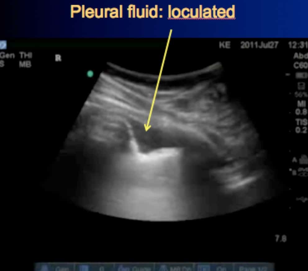
The differential diagnosis of pleural effusion entails consideration of a long list of entities (chart 4.1), 465 but the radiologist should not be discouraged. Peritoneal dialysis/continuous ambulatory peritoneal dialysis. Most pleural effusions, whether free flowing or loculated, are hypoechoic with a sharp echogenic line that delineates the visceral pleura and lung. Pleural effusion is suspected in patients with pleuritic pain, unexplained dyspnea, or suggestive signs. Ultrasound is a reliable method for detecting small pleural effusions and for guiding thoracentesis.
Neutrophilic Loculated Tuberculous Pleural Effusion Incidence Characteristics And Differentiation From Complicated Parapneumonic Effusion The American Journal Of The Medical Sciences from els-jbs-prod-cdn.jbs.elsevierhealth.com .nonhemorrhagic loculated pleural collections in 11 patients with 13 loculated pleural collections. The indication for diagnostic thoracentesis is the new finding of a pleural effusion. Pleural effusion is not a disease. Case contributed by dr prashant mudgal. Pleural effusion is suspected in patients with pleuritic pain, unexplained dyspnea, or suggestive signs. In the latter, there is typically a small amount of fluid. Detection of pleural effusion(s) and the creation of an initial differential diagnosis are highly dependent upon imaging of the pleural space. Pleural effusion is suspected in patients with pleuritic pain, unexplained dyspnea, or suggestive signs.
Process and confirming the diagnosis.
Loculated pleural effusion / pleural effusion is an accumulation of fluid in the pleural cavity between the lining of the lungs and the thoracic cavity. More than one half of these massive. Ultrasound is a reliable method for detecting small pleural effusions and for guiding thoracentesis. Diagnostic tests are indicated to document the presence of pleural fluid and to determine its cause (see figure diagnosis of pleural effusion). Considerations in the differential diagnosis of transudative pleural effusion include the following: Loculated effusions are collections of fluid trapped by pleural adhesions or within pulmonary fissures. Specific differential diagnosis problems generated by pleural nodules, where the main concern is establishing if the complementary ct scan is needed. .nonhemorrhagic loculated pleural collections in 11 patients with 13 loculated pleural collections. The approach to the differential diagnosis of pleural or pericardial effusion starts out with confirmation of these effusions as exudative. Normally, there is a similar retractile force applied to the entire pleural space by adjacent lung. Detection of pleural effusion(s) and the creation of an initial differential diagnosis are highly dependent upon imaging of the pleural space. In the latter, there is typically a small amount of fluid. Pleural effusion is not a disease.
Detection of pleural effusion (s) and the creation of an initial differential diagnosis are highly dependent upon conventional chest radiography and computed tomography (ct) scanning are the primary imaging loculated pleural effusion. Case contributed by dr prashant mudgal.
Share :
Post a Comment
for "Loculated Pleural Effusion Differential Diagnosis / Transcatheter Intrapleural Urokinase For Loculated Pleural Effusion"


Post a Comment for "Loculated Pleural Effusion Differential Diagnosis / Transcatheter Intrapleural Urokinase For Loculated Pleural Effusion"